共2页/22条首页上一页12下一页尾页
回复:22 阅读:4719
Ovarian melanoma (cqz 4)

cqzhao 离线

帖子:5518
粉蓝豆:1071
经验:5755
注册时间:2008-09-29
加关注  |  发消息
楼主 发表于 2009-02-07 01:23|举报|关注(0)
浏览排序[ 顺序 逆序 楼主 支持 精彩 ]  快捷回复
姓    名: ××× 性别:   年龄:  
标本名称:  
简要病史:  
肉眼检查:  

See an interesting case and share with you.

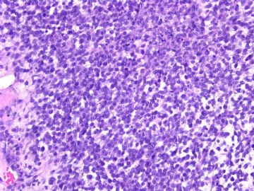
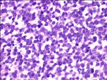
women/about 40 years with a left ovarian mass 13 cm.

Procedure: BSO, hysterectomy, lymph node resection, omentectomy, appendectomy.

Cytomorphology of the tumor is as above photos.

Your interpretations.

  • Ovarian melanoma (cqz 4)图1
    图1
  • Ovarian melanoma (cqz 4)图2
    图2
标签:
本帖最后由 于 2009-02-25 03:02:00 编辑
0
添加参考诊断
×参考诊断
  

cqzhao 离线

帖子:5518
粉蓝豆:1071
经验:5755
注册时间:2008-09-29
加关注  |  发消息
1 楼    发表于2009-02-10 09:45:00举报|引用
返回顶部 | 快捷回复
 inhibin, nse negative
0
回复

Urbino 离线

帖子:66
粉蓝豆:1
经验:68
注册时间:2008-07-27
加关注  |  发消息
2 楼    发表于2009-02-10 12:48:00举报|引用
返回顶部 | 快捷回复
本帖最后由 于 2009-02-10 13:23:00 编辑

 small round blue cells with scant cytoplasm. Dix include

ovarian small cell carcinoma of hypercalcemic type OSCCHT (hyypercalcaemia, EMA, CK focal positive, WT1 diffuse positive)

neuroendocrine small cell carcinoma

lymphoma

melanoma

I think sex-cord stroal tumor could be ruled out.

0
回复

漫游人 离线

帖子:15
粉蓝豆:1
经验:15
注册时间:2009-01-17
加关注  |  发消息
3 楼    发表于2009-02-20 22:08:00举报|引用
返回顶部 | 快捷回复
Based on H&E, I would consider following differentials
1. small cell carcinoma, hypercalcemic type. However, it is unlikely the diagnosis since epithelial markers are all negative.
2. PNET.  CD99 should be positive in most cases. I noticed the current case is negative. 
3. endometrioid stromal sarcoma. There are no characteristic vessels in this case as seen in typical stromal sarcoma of endometrium.
4. melanoma. it can look like anything. IHC can easily rule in or out it.
5. lymphoblastic lymphoma/leukemia. Mitosis is not high, IHC results do not support.
6. rhabdomyosarcoma. Although there is no idenitifiable rhadomyoblast and no IHC result, I still think this should be the favoured diagnosis.
Thank you for the many excellent cases and great effort.

0
回复

cq1981 离线

帖子:336
粉蓝豆:241
经验:395
注册时间:2007-08-28
加关注  |  发消息
4 楼    发表于2009-02-11 16:32:00举报|引用
返回顶部 | 快捷回复
 仔细看细胞核似乎有核沟,猜一个,Langerhans细胞增生症。
0
回复
signature
天将降大任于斯人也,必先苦其心志,劳其筋骨,饿其体肤,空乏其身。

sjp 离线

帖子:954
粉蓝豆:554
经验:965
注册时间:2008-04-28
加关注  |  发消息
5 楼    发表于2009-02-11 17:43:00举报|引用
返回顶部 | 快捷回复
 粒层细胞瘤
0
回复

cqzhao 离线

帖子:5518
粉蓝豆:1071
经验:5755
注册时间:2008-09-29
加关注  |  发消息
6 楼    发表于2009-02-12 11:06:00举报|引用
返回顶部 | 快捷回复
以下是引用sjp在2009-2-11 17:43:00的发言:

 粒层细胞瘤

But inhibin and calretinin are negative.
0
回复

静静鱼 离线

帖子:91
粉蓝豆:3
经验:404
注册时间:2008-06-26
加关注  |  发消息
7 楼    发表于2009-02-12 19:26:00举报|引用
返回顶部 | 快捷回复
 颗粒细胞瘤
0
回复

cqzhao 离线

帖子:5518
粉蓝豆:1071
经验:5755
注册时间:2008-09-29
加关注  |  发消息
8 楼    发表于2009-02-12 19:51:00举报|引用
返回顶部 | 快捷回复
以下是引用静静鱼在2009-2-12 19:26:00的发言:

 颗粒细胞瘤

But inhibin, CD99 and calretinin are negative. Do you still think it is granulosa cell tumor?
0
回复

cqzhao 离线

帖子:5518
粉蓝豆:1071
经验:5755
注册时间:2008-09-29
加关注  |  发消息
9 楼    发表于2009-02-25 02:53:00举报|引用
返回顶部 | 快捷回复
本帖最后由 于 2009-02-25 03:01:00 编辑

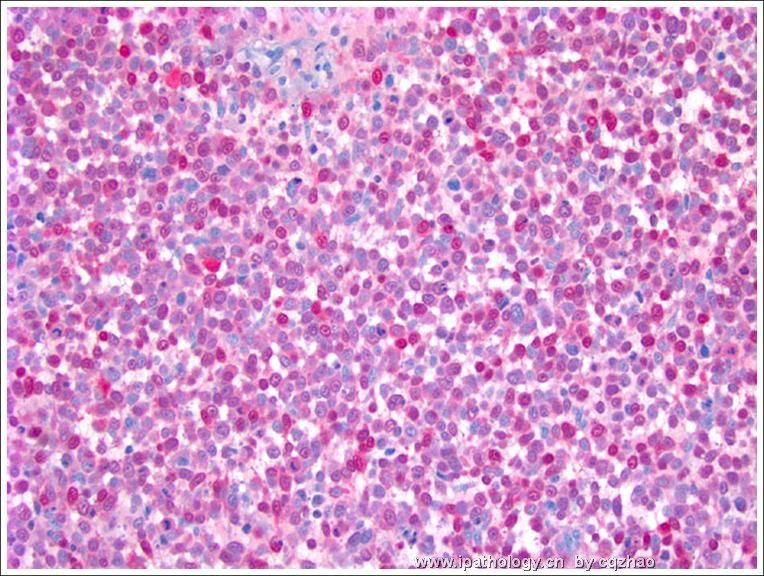
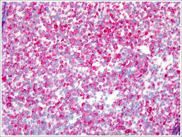
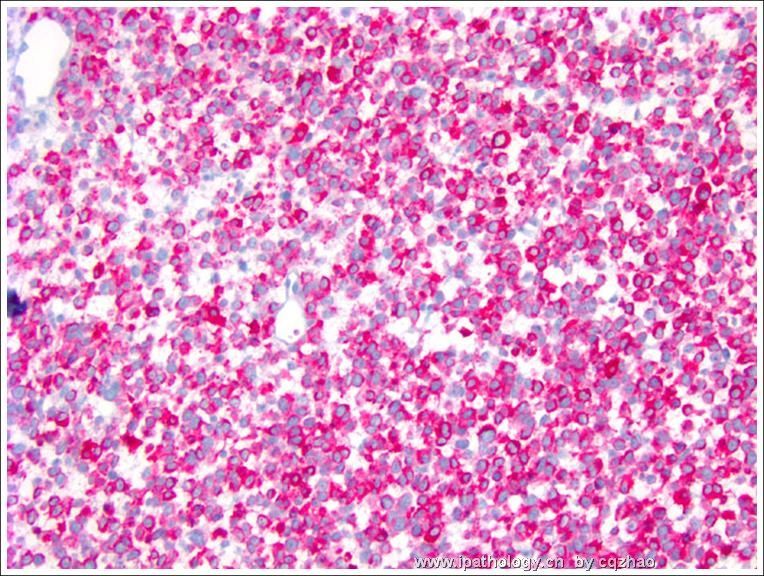
 Stain for s100, HMB 45, Malan-A

This is an easy case. I showed here because it is rare. Clinically the primary lesion have not be found yet. We still think it is a metastatic lesion.  Sometimes we see the metastatic lesions, but cannot find the primary lesions.

Lesson: For unusual cases, always have the differential diagnoses, and then do some IHC to rule out or rule in.

Thank you for reading the case.

cz


名称:图1
描述:图1
名称:图2
描述:图2
名称:图3
描述:图3
0
回复

cqzhao 离线

帖子:5518
粉蓝豆:1071
经验:5755
注册时间:2008-09-29
加关注  |  发消息
10 楼    发表于2009-02-14 19:57:00举报|引用
返回顶部 | 快捷回复
以下是引用cq1981在2009-2-11 16:32:00的发言:

 仔细看细胞核似乎有核沟,猜一个,Langerhans细胞增生症。

IHC results do not support
0
回复

cq1981 离线

帖子:336
粉蓝豆:241
经验:395
注册时间:2007-08-28
加关注  |  发消息
11 楼    发表于2009-02-14 21:33:00举报|引用
返回顶部 | 快捷回复
 排除了粒层细胞瘤、小细胞癌,大部分淋巴瘤,那考虑是低分化的支持-间质细胞瘤或者转移至卵巢的粒细胞肉瘤。
0
回复
signature
天将降大任于斯人也,必先苦其心志,劳其筋骨,饿其体肤,空乏其身。

cqzhao 离线

帖子:5518
粉蓝豆:1071
经验:5755
注册时间:2008-09-29
加关注  |  发消息
12 楼    发表于2009-02-14 22:41:00举报|引用
返回顶部 | 快捷回复
以下是引用cq1981在2009-2-14 21:33:00的发言:

 排除了粒层细胞瘤、小细胞癌,大部分淋巴瘤,那考虑是低分化的支持-间质细胞瘤或者转移至卵巢的粒细胞肉瘤。

What are other differential diagnoses?
0
回复

cq1981 离线

帖子:336
粉蓝豆:241
经验:395
注册时间:2007-08-28
加关注  |  发消息
13 楼    发表于2009-02-17 09:11:00举报|引用
返回顶部 | 快捷回复

还有子宫内膜样间质肉瘤、未分化癌吧,哎,黔驴技穷啊!

0
回复
signature
天将降大任于斯人也,必先苦其心志,劳其筋骨,饿其体肤,空乏其身。

cqzhao 离线

帖子:5518
粉蓝豆:1071
经验:5755
注册时间:2008-09-29
加关注  |  发消息
14 楼    发表于2009-02-17 10:45:00举报|引用
返回顶部 | 快捷回复
 No one can make dx by H&E only for this kind of cases. Should have differential dx first, then order the IHC. Rule in or out based on the IHC results.
0
回复

cqzhao 离线

帖子:5518
粉蓝豆:1071
经验:5755
注册时间:2008-09-29
加关注  |  发消息
15 楼    发表于2009-02-07 07:13:00举报|引用
返回顶部 | 快捷回复
 More history: Pt had no any previous malignant history. She presented with vaginal bleeding. U/S indicated the adanexal mass. Clinical examination and imaging did not find other abnormality.
0
回复

小狼 离线

帖子:37
粉蓝豆:1
经验:46
注册时间:2008-02-27
加关注  |  发消息
16 楼    发表于2009-02-07 23:08:00举报|引用
返回顶部 | 快捷回复
 lympoma?
0
回复

cqzhao 离线

帖子:5518
粉蓝豆:1071
经验:5755
注册时间:2008-09-29
加关注  |  发消息
17 楼    发表于2009-02-08 06:57:00举报|引用
返回顶部 | 快捷回复
以下是引用天山望月在2009-2-7 22:02:00的发言:

 谢谢赵博士分享有趣的病例!估计不易诊断。

我试着描述一下:瘤细胞密集弥漫分布,有点网状连接,隐约可见 Call-Exner小体样结构,细胞小圆形,界不清,核圆形,有核仁,偶见核沟,高核分裂像。

考虑较多:1、卵巢成年型粒层细胞瘤(弥漫型)?2、间质肉瘤?3、PNET?4、淋巴瘤?(可见浆细胞)

5、类癌?等,免疫组化有帮助。

切除部位较多,估计病变复杂。期待更多讨论。。。

Only lesion was in this overy. All other orgains showed no significant lesions.
0
回复

cqzhao 离线

帖子:5518
粉蓝豆:1071
经验:5755
注册时间:2008-09-29
加关注  |  发消息
18 楼    发表于2009-02-09 03:00:00举报|引用
返回顶部 | 快捷回复
 Epithelial markers, CD45, CD3, Cd20 are negative
0
回复

zchzmf 离线

帖子:701
粉蓝豆:17
经验:864
注册时间:2007-03-15
加关注  |  发消息
19 楼    发表于2009-02-09 09:57:00举报|引用
返回顶部 | 快捷回复
 CD99  vim  NSE   抑制素
0
回复

fuying 离线

帖子:362
粉蓝豆:2
经验:390
注册时间:2008-04-10
加关注  |  发消息
20 楼    发表于2009-02-09 10:28:00举报|引用
返回顶部 | 快捷回复
 卵巢成年型粒层细胞瘤
0
回复
回复:22 阅读:4719
共2页/22条首页上一页12下一页尾页
【免责声明】讨论内容仅作学术交流之用,不作为诊疗依据,由此而引起的法律问题作者及本站不承担任何责任。
快速回复
进入高级回复
您最多可输入10000个汉字,按 "Ctrl" + "Enter" 直接发送
搜索回复/乘电梯 ×
按内容
按会员
乘电梯
合作伙伴
友情链接