| 图片: | |
|---|---|
| 名称: | |
| 描述: | |
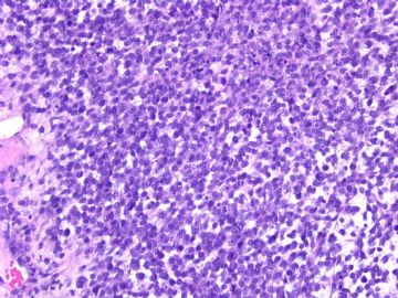
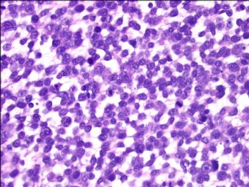
- Ovarian melanoma (cqz 4)
| 姓 名: | ××× | 性别: | 年龄: | ||
| 标本名称: | |||||
| 简要病史: | |||||
| 肉眼检查: | |||||
See an interesting case and share with you.
women/about 40 years with a left ovarian mass 13 cm.
Procedure: BSO, hysterectomy, lymph node resection, omentectomy, appendectomy.
Cytomorphology of the tumor is as above photos.
Your interpretations.
-
本帖最后由 于 2009-02-25 03:02:00 编辑
-
本帖最后由 于 2009-02-25 03:01:00 编辑
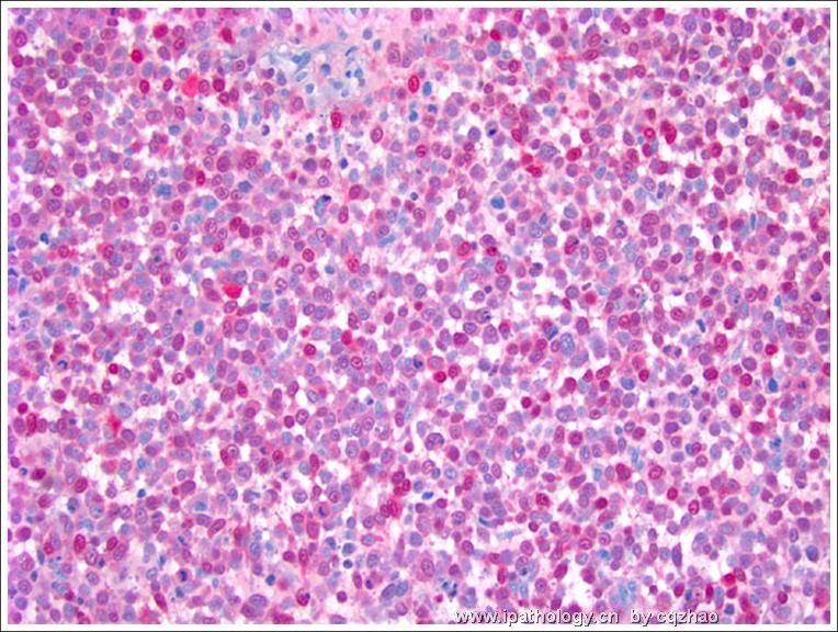
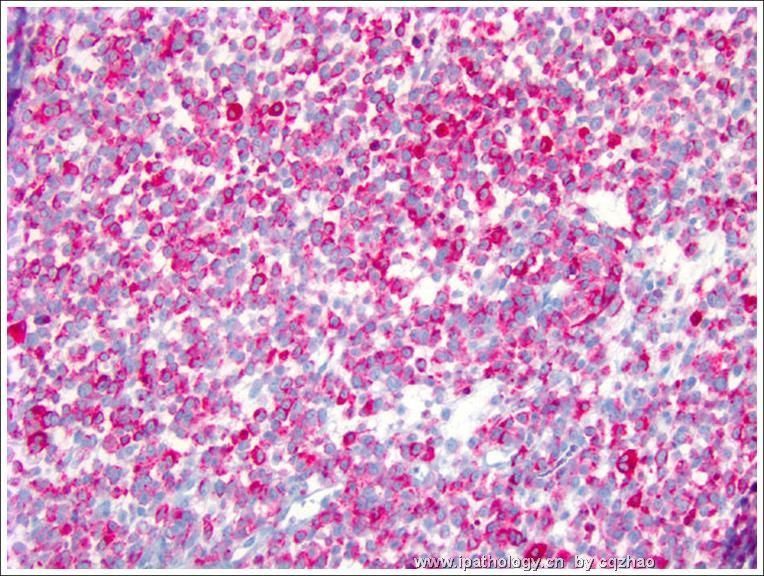
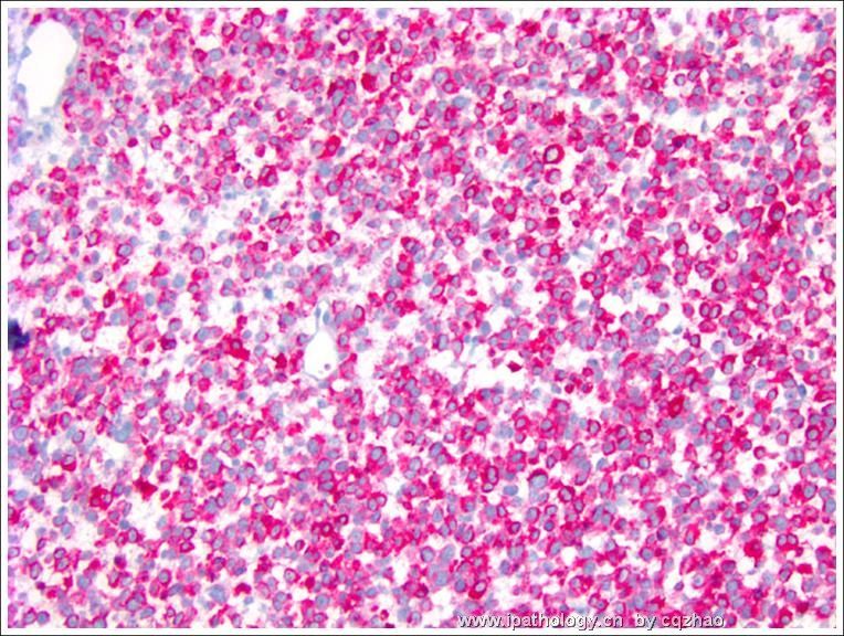
Stain for s100, HMB 45, Malan-A
This is an easy case. I showed here because it is rare. Clinically the primary lesion have not be found yet. We still think it is a metastatic lesion. Sometimes we see the metastatic lesions, but cannot find the primary lesions.
Lesson: For unusual cases, always have the differential diagnoses, and then do some IHC to rule out or rule in.
Thank you for reading the case.
cz
名称:图1
描述:图1
名称:图2
描述:图2
名称:图3
描述:图3
| 以下是引用漫游人在2009-2-20 22:08:00的发言: Based on H&E, I would consider following differentials |
-
Based on H&E, I would consider following differentials
1. small cell carcinoma, hypercalcemic type. However, it is unlikely the diagnosis since epithelial markers are all negative.
2. PNET. CD99 should be positive in most cases. I noticed the current case is negative.
3. endometrioid stromal sarcoma. There are no characteristic vessels in this case as seen in typical stromal sarcoma of endometrium.
4. melanoma. it can look like anything. IHC can easily rule in or out it.
5. lymphoblastic lymphoma/leukemia. Mitosis is not high, IHC results do not support.
6. rhabdomyosarcoma. Although there is no idenitifiable rhadomyoblast and no IHC result, I still think this should be the favoured diagnosis.
Thank you for the many excellent cases and great effort.
-
本帖最后由 于 2009-02-10 13:23:00 编辑
small round blue cells with scant cytoplasm. Dix include
ovarian small cell carcinoma of hypercalcemic type OSCCHT (hyypercalcaemia, EMA, CK focal positive, WT1 diffuse positive)
neuroendocrine small cell carcinoma
lymphoma
melanoma
I think sex-cord stroal tumor could be ruled out.
| 以下是引用天山望月在2009-2-7 22:02:00的发言:
谢谢赵博士分享有趣的病例!估计不易诊断。 我试着描述一下:瘤细胞密集弥漫分布,有点网状连接,隐约可见 Call-Exner小体样结构,细胞小圆形,界不清,核圆形,有核仁,偶见核沟,高核分裂像。 考虑较多:1、卵巢成年型粒层细胞瘤(弥漫型)?2、间质肉瘤?3、PNET?4、淋巴瘤?(可见浆细胞) 5、类癌?等,免疫组化有帮助。 切除部位较多,估计病变复杂。期待更多讨论。。。 |
