| 图片: | |
|---|---|
| 名称: | |
| 描述: | |
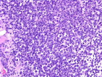
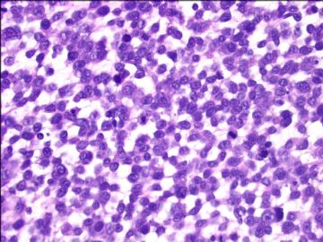
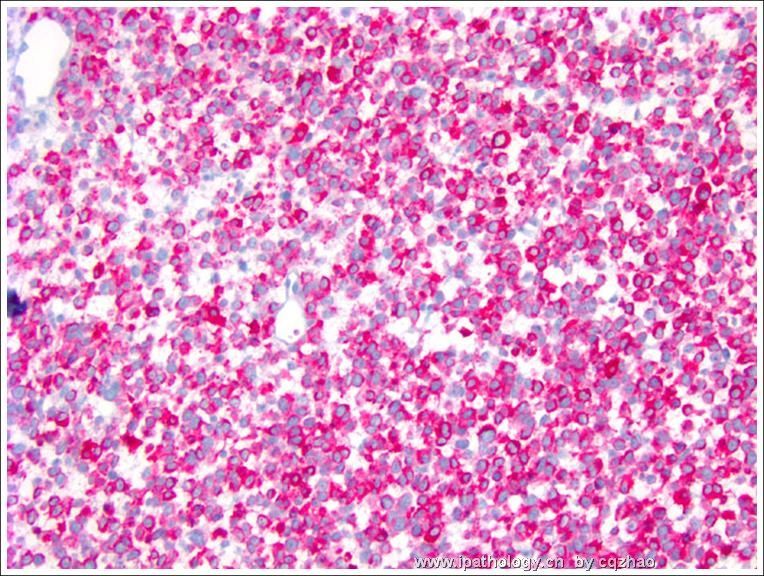
- Ovarian melanoma (cqz 4)
| 姓 名: | ××× | 性别: | 年龄: | ||
| 标本名称: | |||||
| 简要病史: | |||||
| 肉眼检查: | |||||
See an interesting case and share with you.
women/about 40 years with a left ovarian mass 13 cm.
Procedure: BSO, hysterectomy, lymph node resection, omentectomy, appendectomy.
Cytomorphology of the tumor is as above photos.
Your interpretations.
-
本帖最后由 于 2009-02-25 03:02:00 编辑
| 以下是引用漫游人在2009-2-20 22:08:00的发言: Based on H&E, I would consider following differentials |
-
本帖最后由 于 2009-02-25 03:01:00 编辑
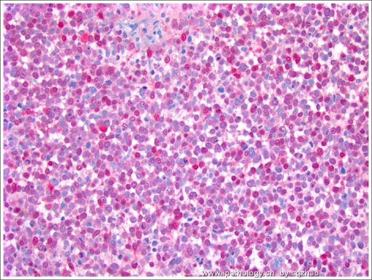
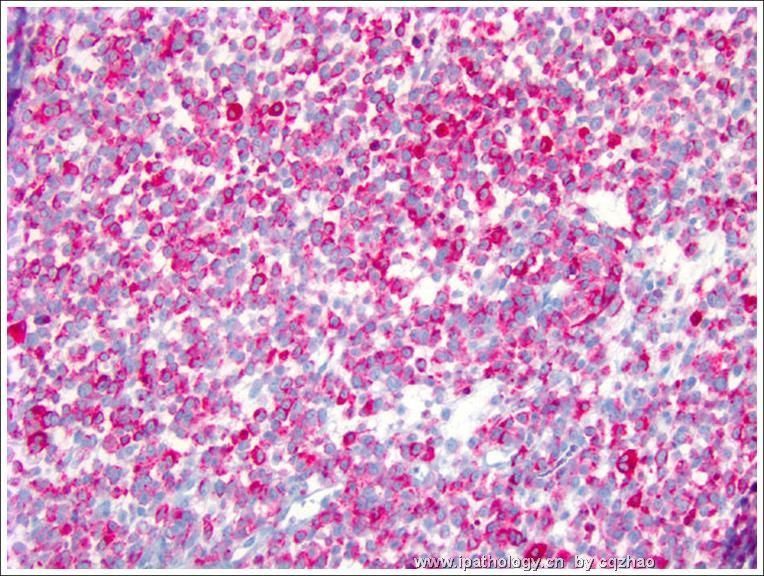
Stain for s100, HMB 45, Malan-A
This is an easy case. I showed here because it is rare. Clinically the primary lesion have not be found yet. We still think it is a metastatic lesion. Sometimes we see the metastatic lesions, but cannot find the primary lesions.
Lesson: For unusual cases, always have the differential diagnoses, and then do some IHC to rule out or rule in.
Thank you for reading the case.
cz
名称:图1
描述:图1
名称:图2
描述:图2
名称:图3
描述:图3
