共1页/12条首页上一页1下一页尾页
回复:12 阅读:2761
B1759乳腺肿块

全子 离线

帖子:3318
粉蓝豆:74
经验:3546
注册时间:2006-10-19
加关注  |  发消息
楼主 发表于 2009-01-20 08:57|举报|关注(0)
浏览排序[ 顺序 逆序 楼主 支持 精彩 ]  快捷回复
姓    名: ××× 性别:  女 年龄:  39
标本名称:  乳腺肿块
简要病史:  发现肿块3月
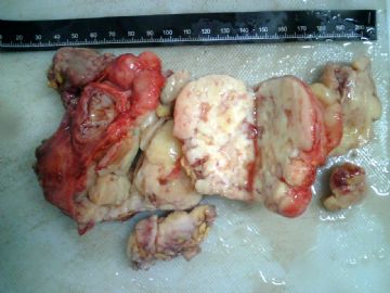
肉眼检查:  10cm分叶状肿块
  • 乳腺肿块图1
    图1
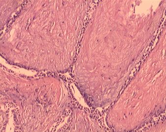
  • 乳腺肿块图2
    图2
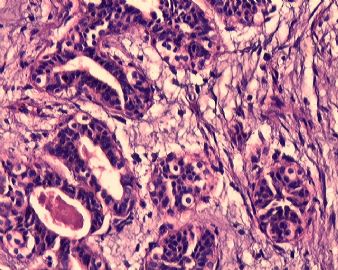
  • 乳腺肿块图3
    图3
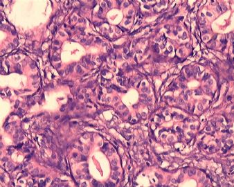
  • 乳腺肿块图4
    图4
  • 乳腺肿块图5
    图5
  • 乳腺肿块图6
    图6
  • 乳腺肿块图7
    图7
  • 乳腺肿块图8
    图8
  • 乳腺肿块图9
    图9
  • 乳腺肿块图10
    图10
  • 乳腺肿块图11
    图11
标签:叶状肿瘤
0
添加参考诊断
×参考诊断
  

全子 离线

帖子:3318
粉蓝豆:74
经验:3546
注册时间:2006-10-19
加关注  |  发消息
1 楼    发表于2009-01-20 09:01:00举报|引用
返回顶部 | 快捷回复
本帖最后由 于 2009-01-20 09:13:00 编辑

好多上不去的,再传

  • 图1
  • 图2
  • 图3
  • 图4
  • 图5
  • 图6
  • 图7
  • 图8
  • 图9
  • 图10
0
回复

风的影子 离线

帖子:4277
粉蓝豆:3125
经验:8142
注册时间:2006-10-18
加关注  |  发消息
2 楼    发表于2009-01-20 16:27:00举报|引用
返回顶部 | 快捷回复
本帖最后由 于 2009-01-20 21:40:00 编辑

 1\纤维腺瘤伴导管内上皮不典型增生,

  2\考虑交界性叶状肿瘤

0
回复

cqzhao 离线

帖子:5518
粉蓝豆:1071
经验:5755
注册时间:2008-09-29
加关注  |  发消息
3 楼    发表于2009-01-20 17:02:00举报|引用
返回顶部 | 快捷回复

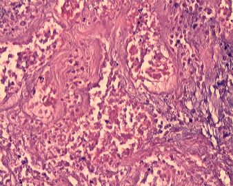
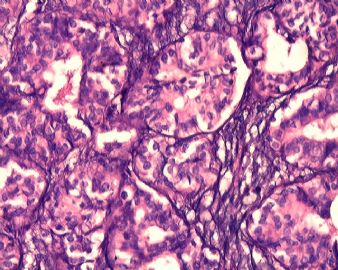
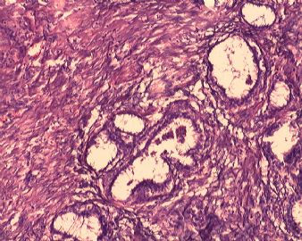
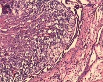
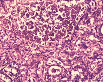
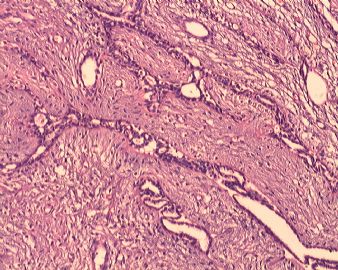
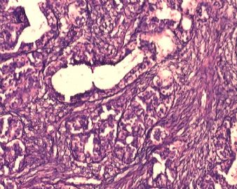
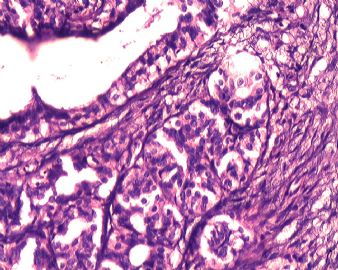
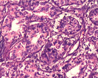
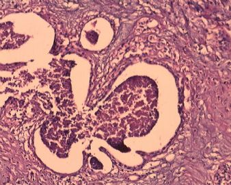
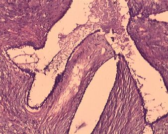
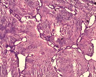
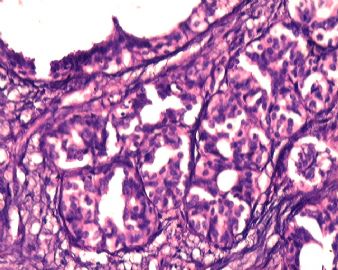
 it is a complicated case. 3 month, 10 cm, heterogeneous growth in gross photos. It iooks like a fibroepithelial tumor with focal adenosis and necrosis. Glandular atypia is not noted. Focal leaf-like structures are present. I would classify the tumor as phyllodes tumor.  You need to check stroma atypia, mitosis, and margins carefully. I did not see mitosis and obvious stroma atypia from above photos. WHO classification for phyllodes: benign, borderlin, malignant. I favor a benign phyllodes. Someetimes fibroadenoma and benign phyllodes are difficult to distinguish. Some people use the term fibroadenoma-phyllodes for some lesion. Large fibroaenomas can have necrosis also. Anyway I would call phyllodes but not fibroadenoma based on the clinical presentation and size. The treatment should wide local excision. it means keep wide clear margins.

The reveiw sof photos and true slides are different. Jus for your reference.

Thank for sharing interesting case and the beautiful photos.

cz

0
回复

小鹤 离线

帖子:206
粉蓝豆:80
经验:1321
注册时间:2007-06-10
加关注  |  发消息
4 楼    发表于2009-01-20 17:04:00举报|引用
返回顶部 | 快捷回复
 诊断 叶状肿瘤似乎条件不够!!!----纤维腺瘤
0
回复
signature
好好学习,天天向上

全子 离线

帖子:3318
粉蓝豆:74
经验:3546
注册时间:2006-10-19
加关注  |  发消息
5 楼    发表于2009-01-20 20:29:00举报|引用
返回顶部 | 快捷回复

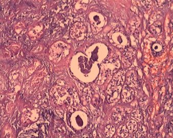
 1.非常赞成赵老师的分析,从大体和局部的镜下所见看,我觉得诊断叶状肿瘤是没问题的,根据间质的情况倾向良性

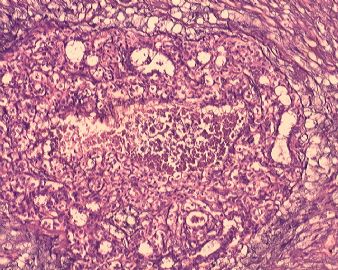
2.本例疑惑的是导管内的坏死,想考虑导管内癌了,但是就是觉得形态还不够,能不能描写不典型增生伴有坏死

感谢赵老师!

0
回复

wangdingding 离线

帖子:1474
粉蓝豆:98
经验:6042
注册时间:2006-10-19
加关注  |  发消息
6 楼    发表于2009-01-20 20:50:00举报|引用
返回顶部 | 快捷回复
 叶状肿瘤伴小叶增生?
0
回复

cqzhao 离线

帖子:5518
粉蓝豆:1071
经验:5755
注册时间:2008-09-29
加关注  |  发消息
7 楼    发表于2009-01-20 22:32:00举报|引用
返回顶部 | 快捷回复
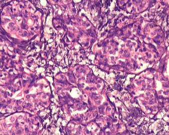
 全子 : When you talk atypical proliferation you mean glandular component. I cannot apprecitate the atypia from your photos. No DCIS is noted. You can write something in your comment. Focal adenosis and ductal hyperplasia show central necrosis with mild cytologic atypia. It is difficult to evaluate the nature of atypia due to the necrosis. Of cause if you feel confident in your glass slide that atpia is present you can write in your diagnosis line.  Anyway I did not see glandular atypia in the photos. In other word if you call benign phyllodes the patient should have close follow up. The treatment will not have any change even if you call focal glandular atypia.  for u reference.
0
回复

cqzhao 离线

帖子:5518
粉蓝豆:1071
经验:5755
注册时间:2008-09-29
加关注  |  发消息
8 楼    发表于2009-01-20 23:06:00举报|引用
返回顶部 | 快捷回复
 Necrosis can be present in many benign conditions, such as, sclerosing adenosis, UDH, intraductal papilloma, apocrine metaplasia .........
0
回复

全子 离线

帖子:3318
粉蓝豆:74
经验:3546
注册时间:2006-10-19
加关注  |  发消息
9 楼    发表于2009-01-22 22:03:00举报|引用
返回顶部 | 快捷回复
 谢谢赵老师,我就是这样描写并建议密切随访的
0
回复

病理小精灵 离线

帖子:78
粉蓝豆:734
经验:85
注册时间:2009-01-12
加关注  |  发消息
10 楼    发表于2009-01-22 23:16:00举报|引用
返回顶部 | 快捷回复

间质增生很厉害,要数核分裂!

诊断:叶状肿瘤伴导管上皮不典型增生。

0
回复

海浪信使 离线

帖子:1547
粉蓝豆:161
经验:2339
注册时间:2006-09-26
加关注  |  发消息
11 楼    发表于2009-01-23 14:44:00举报|引用
返回顶部 | 快捷回复
 良性叶状肿瘤应该没有问题。导管上皮的增生也没有问题,虽然看起来挺可怕的样子(不除外外源性激素造成的,如某些保健食品或药品及化妆品等,这个年纪的女性还是很注意保养自己的哈),但是两型上皮还是存在的,细胞的异型性也不明显。难以解释的是坏死,查了几本书也没找到好的解释。期待老师们的讲解。
0
回复
signature
当你有选择的时候,不是选择正确的,而是选择不让你后悔的!

abin 离线

帖子:7416
粉蓝豆:6014
经验:11787
注册时间:2006-10-08
加关注  |  发消息
12 楼    发表于2009-01-24 12:52:00举报|引用
返回顶部 | 快捷回复

 大体图像是分叶状肿瘤,镜下照片看不清。似乎间质增生,导管也增生。

需要改进制片质量哦

0
回复
signature

华夏病理/粉蓝医疗

为基层医院病理科提供全面解决方案,

努力让人人享有便捷准确可靠的病理诊断服务。


回复:12 阅读:2761
共1页/12条首页上一页1下一页尾页
【免责声明】讨论内容仅作学术交流之用,不作为诊疗依据,由此而引起的法律问题作者及本站不承担任何责任。
快速回复
进入高级回复
您最多可输入10000个汉字,按 "Ctrl" + "Enter" 直接发送
搜索回复/乘电梯 ×
按内容
按会员
乘电梯
合作伙伴
友情链接