


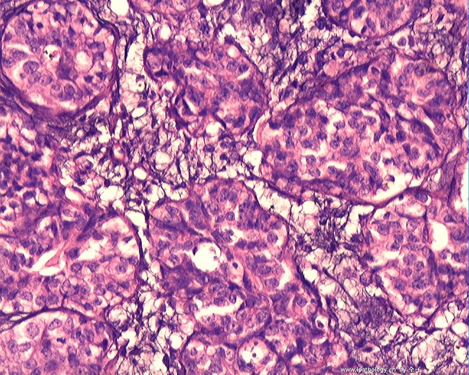
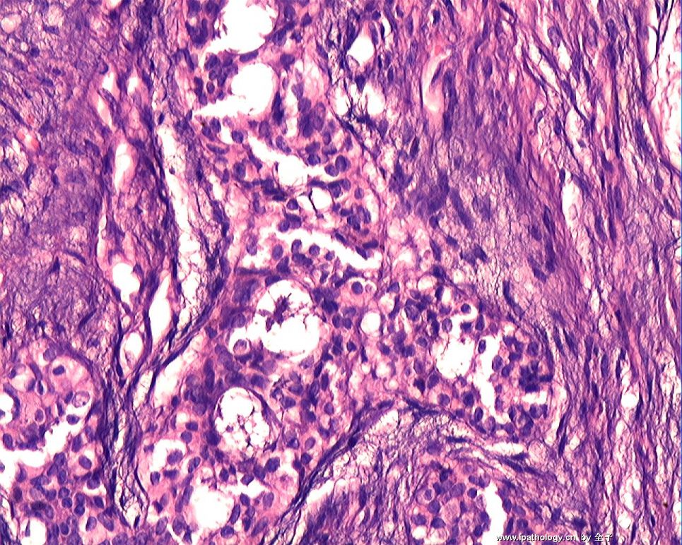


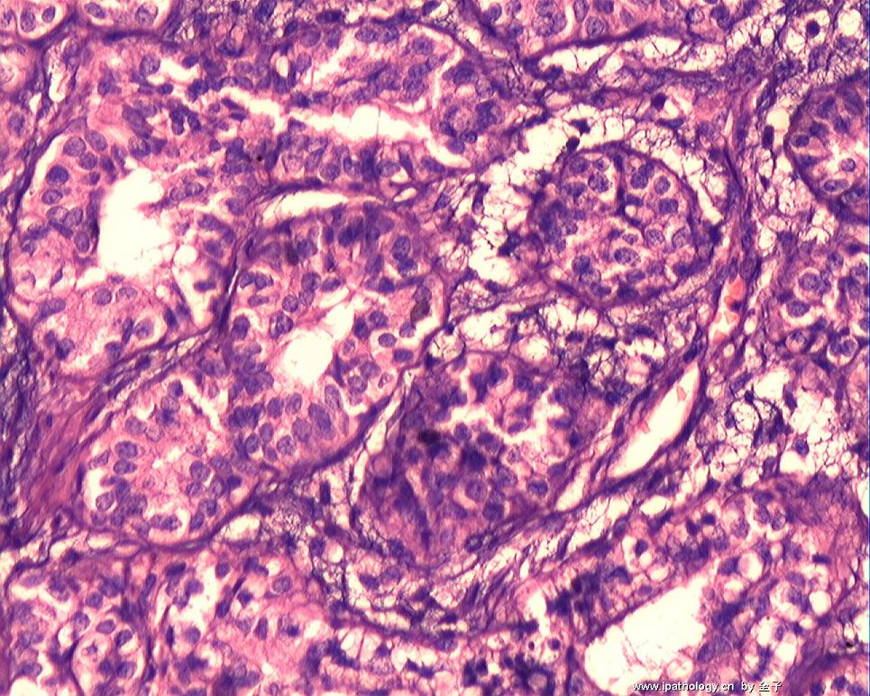
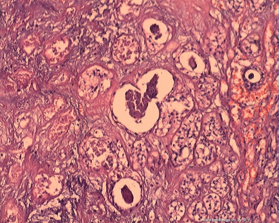
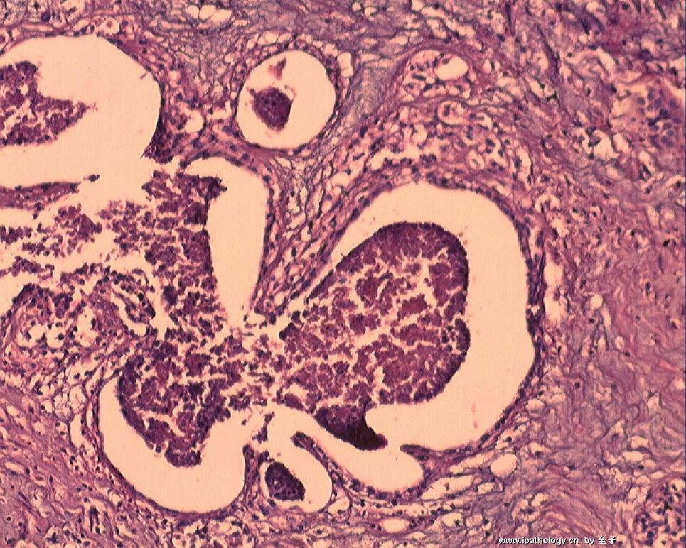
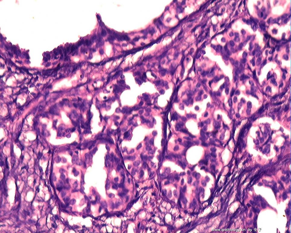


 ),但是两型上皮还是存在的,细胞的异型性也不明显。难以解释的是坏死,查了几本书也没找到好的解释。期待老师们的讲解。
| 图片: | |
|---|---|
| 名称: | |
| 描述: | |
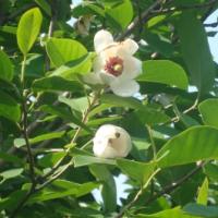
- B1759乳腺肿块
 图1
图1 图2
图2 图3
图3 图4
图4 图5
图5 图6
图6 图7
图7 图8
图8 图9
图9 图10
图10 图11
图11
| 姓 名: | ××× | 性别: | 女 | 年龄: | 39 |
| 标本名称: | 乳腺肿块 | ||||
| 简要病史: | 发现肿块3月 | ||||
| 肉眼检查: | 10cm分叶状肿块 | ||||
标签:叶状肿瘤
相关帖子
×参考诊断
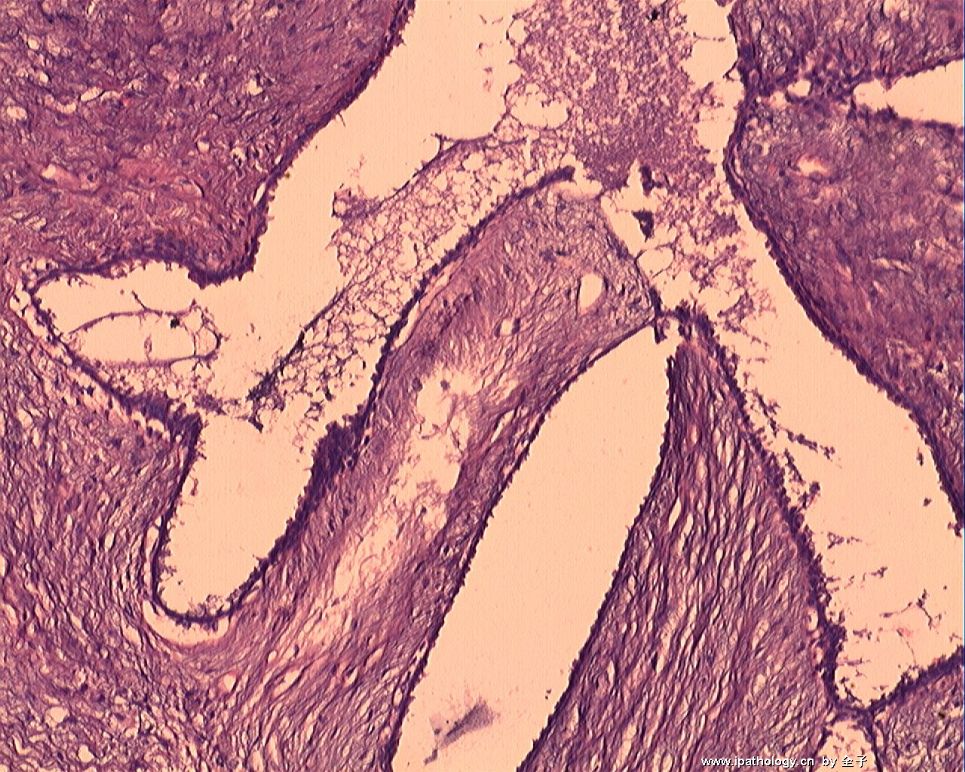
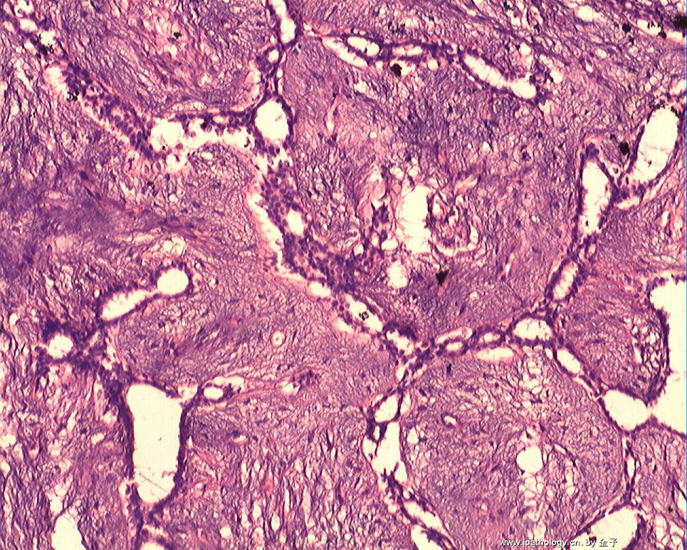
it is a complicated case. 3 month, 10 cm, heterogeneous growth in gross photos. It iooks like a fibroepithelial tumor with focal adenosis and necrosis. Glandular atypia is not noted. Focal leaf-like structures are present. I would classify the tumor as phyllodes tumor. You need to check stroma atypia, mitosis, and margins carefully. I did not see mitosis and obvious stroma atypia from above photos. WHO classification for phyllodes: benign, borderlin, malignant. I favor a benign phyllodes. Someetimes fibroadenoma and benign phyllodes are difficult to distinguish. Some people use the term fibroadenoma-phyllodes for some lesion. Large fibroaenomas can have necrosis also. Anyway I would call phyllodes but not fibroadenoma based on the clinical presentation and size. The treatment should wide local excision. it means keep wide clear margins.
The reveiw sof photos and true slides are different. Jus for your reference.
Thank for sharing interesting case and the beautiful photos.
cz
-
wangdingding 离线
- 帖子:1474
- 粉蓝豆:98
- 经验:6042
- 注册时间:2006-10-19
- 加关注 | 发消息
-
全子 : When you talk atypical proliferation you mean glandular component. I cannot apprecitate the atypia from your photos. No DCIS is noted. You can write something in your comment. Focal adenosis and ductal hyperplasia show central necrosis with mild cytologic atypia. It is difficult to evaluate the nature of atypia due to the necrosis. Of cause if you feel confident in your glass slide that atpia is present you can write in your diagnosis line. Anyway I did not see glandular atypia in the photos. In other word if you call benign phyllodes the patient should have close follow up. The treatment will not have any change even if you call focal glandular atypia. for u reference.


