


| 图片: | |
|---|---|
| 名称: | |
| 描述: | |
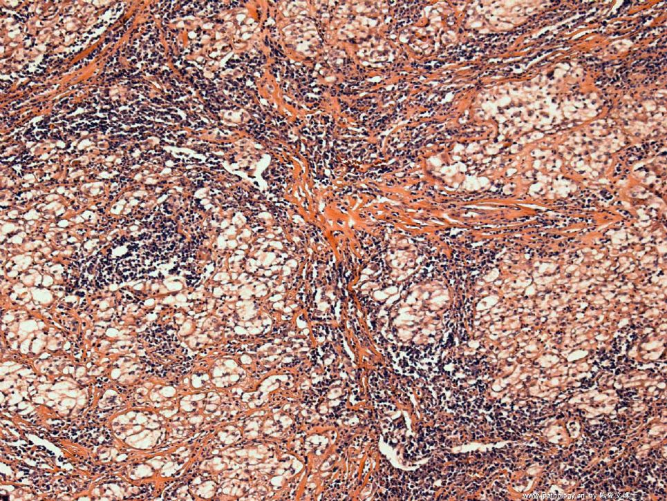
- Anterior Mediastinal FNA
Your questions are good and some require a long answer. If you are going to The Chinese Cytology Meeting this March in Guangzhou, that will be very beneficial to you, Dr. Zhao and I are both going. There will be a pre-conference Thyroid FNA work shop organized by 金域, it will pretty much answer all your questions in detail.
1, yes, thyroid FNA is one of the most common FNA samples seen here at US. I see an average 6-10 thyroid FNA a day here.
2, Well-differentiated follicular carcinoma is generally not a cytologic diagnosis. But, the majority of thyroid carcinoma are papillary carcinoma anyway.
3, you are right.
4, Many colloid nodules can be separated from follicular neoplasms with confident. Some however cannot.
-
本帖最后由 于 2009-02-02 21:25:00 编辑
想请教陈博士和赵博士几个问题,请赐教,谢谢!
1、甲状腺FNA多吗?
2、滤泡性癌诊断准确率怎样?
3、FNA主要依据细胞的异型性,没有组织学结构,而高分化甲状腺癌,需根据生物学行为,即有无浸润包膜或间质等,此类报告难发,不知陈博士和赵博士有何看法?
4、结节性甲状腺肿与腺瘤FNA能区分吗?怎样区分?
呵呵,问题多了点,因为我特喜欢穿刺细胞学,曾做过许多乳腺和甲状腺细针穿刺,想了解一下国外的情况,并期待多发些穿刺的病例,包括典型的或不典型的。
再次感谢!


- 广州金域病理
中国的细胞学病理医生实在是太少了,很多医院包括很多三甲医院都没有专门看细胞学的病理医生,都是看组织学的病理医生兼看的,这样带来了很大的问题就是,看贯了组织片的病理医生是不会重视细胞学的,觉得细胞学很多是术前诊断风险有大,细胞学有不好看,所以很多医生都不是很重视细胞学.如果连病理科都不重视细胞学更何况让医院去重视呢?不过现在越来越多的人认识到了细胞学的重要性,就是因为是术前诊断,所以就更显得细胞学的重要了.不过我还是建议应该让专人学细胞学,做细胞学的专家.毕竟细胞学入门容易,要想提高需要非常丰富的经验积累.现在很多医院的细胞穿刺都是临床来穿的,在把片送到细胞室.个人觉得还是应该让我们自己的细胞学医生来穿刺,好处很多,第一看到了病人,通过交流可以大致了解病情,比临床医生写在申请单上的病情详细多了,第二,通过自己触摸肿块的大小,硬度,边界,可以大致在心中对肿块有个大致的印象,经验丰富后,触摸肿块就可以大致的知道是什么疾病了,第三,如果第一针没有穿到,可以马上穿第二,第三针,为病人节省了很多时间很精力,等等等等.
这只是我对目前我们细胞学的这种氛围一点寓见,说得不对请各位专家批评,
|
译上楼:OK!好, I think that everybody agrees that this is a malignant germ cell tumor. 我想大家都同意为恶性生殖细胞肿瘤,The question is how to subtype it.问题是如何分型? The immunoprofile is not for typical seminoma and it fits more for embrynocarcinoma. 免疫表达不象精原细胞瘤更象是胚胎癌,But, the morphology favors seminoma. 形态象精原细胞瘤。My colleague (he is a very good pathologist)signed this case out as "malignant germ cell tumor, unclassifiable", 我的同事一位非常有能力的病理学家签发报告为末能分类的恶性生殖细胞肿瘤。I am not entirely agree with him. 我并不完全同意他,The ponit is that if the clinical management is no difference, then it should not matter.尽管临床处理没有明显的不同, I also agree with Dr. Zhao that we hope that our Chinese cytopathologists will be more proactive to participate in discussions. 我同意赵老师的意见,希望咱们中国的病理医生们应该积极的参加讨论,We as american trained Chinese pathologists working in the US are usually very busy in our daily diagnostic work and we spend our time here on the web is because we want cytopathology in China will catch up sooner than later. 我们做为受美国训练的华裔病理学家,每天工作非常紧张,花费时间在网上的目的是为了使中国的细胞病理进步更快。Thanks all for participation.感谢所有参加讨论的人。 |
OK! I think that everybody agrees that this is a malignant germ cell tumor. The question is how to subtype it. The immunoprofile is not for typical seminoma and it fits more for embrynocarcinoma. But, the morphology favors seminoma. My colleague (he is a very good pathologist)signed this case out as "malignant germ cell tumor, unclassifiable", I am not entirely agree with him. The ponit is that if the clinical management is no difference, then it should not matter.
I also agree with Dr. Zhao that we hope that our Chinese cytopathologists will be more proactive to participate in discussions. We as american trained Chinese pathologists working in the US are usually very busy in our daily diagnostic work and we spend our time here on the web is because we want cytopathology in China will catch up sooner than later. Thanks all for participation.
| 以下是引用陈隆文博士在2009-1-15 12:16:00的发言: It is negative for PLAP, CD117, and OCT3/4. You still think it is seminoma? |
-
本帖最后由 于 2009-01-14 11:32:00 编辑
赵老师说的对,中国的细针穿刺病理学开展的不算广泛,就算广泛开展也好象并不入主流,许多签发细针穿刺病理报告的或许还不是病理医生,是病理技师,是细胞人员,这是非常不合理的。也是很危险的。根本没有细针穿刺病理系统训练,人人都是自学成才,或者单挑拜师学艺。话说长了,不在此展开,国家花费大量时间大量的金钱培养所谓的博士硕士,搞大量所谓的科研,给祖国医学的宝库里增添一堆堆的垃圾,根本不接触临床病理,毕业后直接当病理高级医生用,非常可怕,非常可恨,非常荒唐,但这些不是我们所能管的事情。
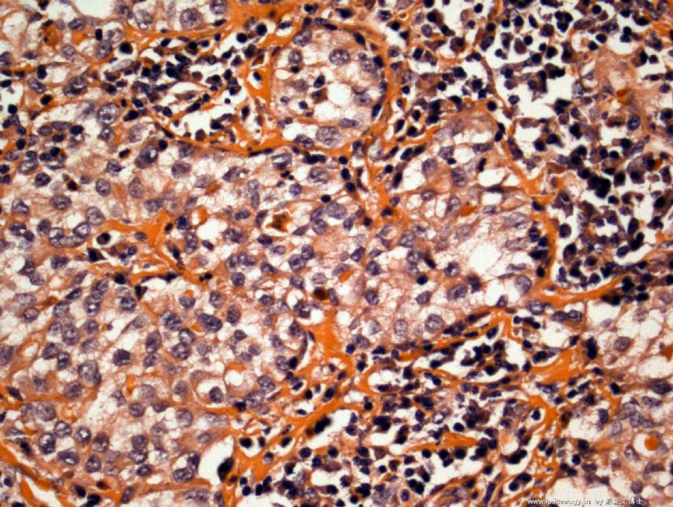
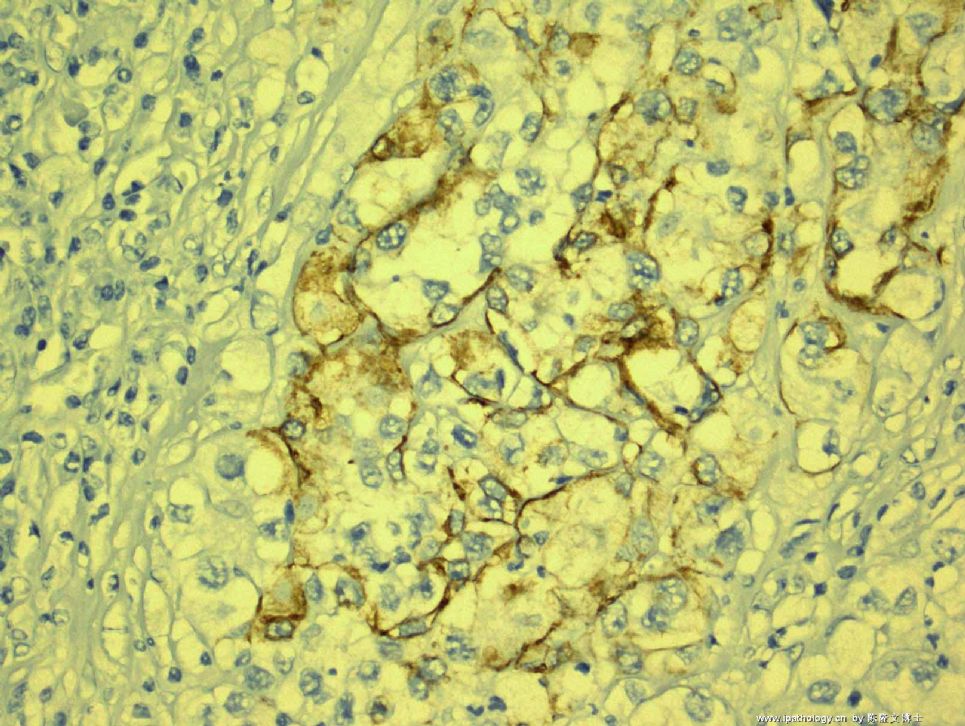
本例有明显的上皮样细胞,AE1/AE3 focally and CD30阳性,还有明显的淋巴样细胞,两种成份,上皮样细胞呈巢状,围绕以淋巴样细胞。考虑生殖细胞肿瘤,精原细胞瘤。
-
本帖最后由 于 2009-01-14 10:58:00 编辑
译上楼:Quickly see the case. 快速阅读本例, It is an excellent teaching case with cytology, surgical and IHC results. Few people are intrerested.该例是非常好的教学病例,但是关注的人极少。Based on the Cytologic surgical and IHC results the first year of residents In the US should know the diagnosis after reviewing the book. 根据细胞学,手术标本活检病理及免疫组化,美国第一年的病理住院医生看过病理教课书之后,都应该了解病理结果The second year of residents should know the diagnosis or at least which category of the tumor . 第二年的病理住院医生应该知道诊断结果,至少应该知道肿瘤的范围。When we have problem in study, we need to check book for help.如果有问题应该及时查书。I assume that most of pathologists in China do not systemically learn FNA cytology or seldom sign out fna cases.可能中国的病理医生没有系统学习过细针穿刺细胞学,很少签发细针穿刺病理报告。 This may be the reason why few people here are not interested in FNA cases. I may be wrong. 这就是为什么很少有人关注细针穿刺的帖子。也许我说的不对。
-
本帖最后由 于 2009-01-14 09:41:00 编辑
Quickly see the case. It is an excellent teaching case with cytology, surgical and IHC results. Few people are intrerested.
Based on the Cytologic surgical and IHC results the first year of residents In the US should know the diagnosis after reviewing the book. The second year of residents should know the diagnosis or at least which category of the tumor .
When we have problem in study, we need to check book for help.
I assume that most of pathologists in China do not systemically learn FNA cytology or seldom sign out fna cases. This may be the reason why few people here are not interested in FNA cases.
I may be wrong.
I am surprised that now I gave you the biopsy and the immunostain results, I did not get much response. At least, I would like to know how you are going to sign-out this case???
Remember, sometimes in pathology, there is no absolute "right" or "wrong" diagnosis, it is very important to think outside the box and get the patient treated appropriately!
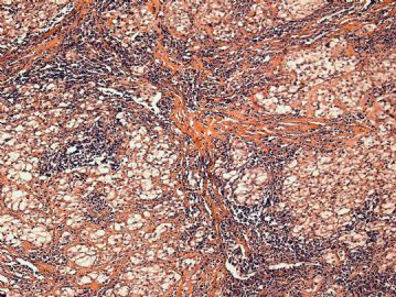 图1
图1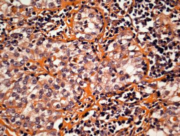 图2
图2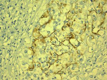 图3
图3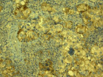 图4
图4
From the cytology, I think that everyone agree that it is malignant. The location of anterior mediatinum will let us think the following differential diagnosis: 1) Thymic Carcinoma; 2) Malignant lymphoma; 3) Malignant germ cell tumor; 4)Malignant thyroid carcinoma. I am glad that you are all considering the above possibility. But, remember that we most likely need immunostains to make a definitive diagnosis and sometimes, cytology has its limitation of not getting enough cells for cell block. So, in this case I asked for a biopsy. My colleague, Dr. Hansel, just brought the biopsy and a LOT OF immunostains to me. I just took some pictures and I am summarizing the immunostains:
Positive results: Only AE1/AE3 focally and CD30
Negative results: CAM5.2, TTF-1, CD20, CD45(LCA), ALK-1, PLAP, CD117,OCT3/4,bcl-2, p53, AFP.
Any thoughts on what this is?


