| 图片: | |
|---|---|
| 名称: | |
| 描述: | |
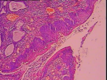
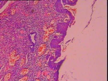
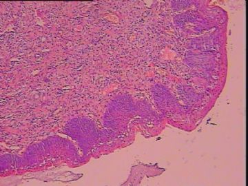
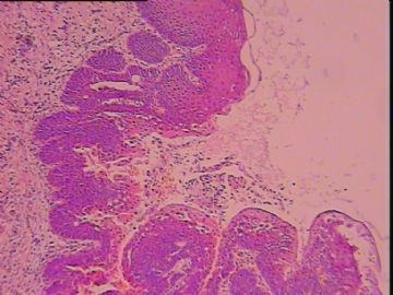
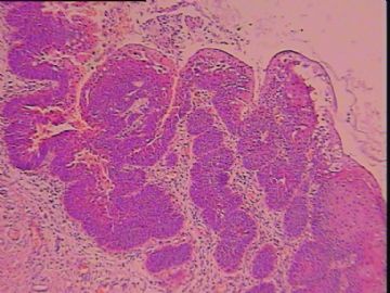
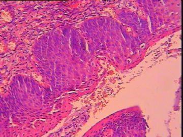
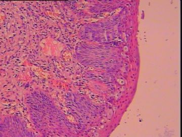
- 65岁,宫颈基底细胞增生?CIN?
In my humble opinion,there is no CIN in this case based on morphology alone. If you pay little attention to the surface change, you will notice the presence of hyperkeratosis and parakeratosis which tends to be mistaken for "HPV infection" both cytologically and histopathologically. So this is a typical case in an old lady with most likely prolapse-related change. The so-called "hyperplastic change at the basal portion" is likely caused by prolase-related stimulation. The p16 immunostain in this case is simply some background staining and should be interpreted as negative. If she has HPV test, my bet is that she is likely HPV-negative. So I would render my diagnosis as follow:
- Benign squamous mucosa with prolapse-related change, negative for dysplasia.

- 不坠青云之志,长怀赤子之心
| 以下是引用cqzhao在2010-9-11 3:21:00的发言: It is difficult case. H&E slides show cervical epithelial proliferation in this 65 y/f. Carefully review the stains for p16 and ki67 and feel that no enough evidence of high grade dysplasia is present. Favor basal cell proliferation. |
支持!
| 以下是引用cqzhao在2010-9-11 3:21:00的发言: It is difficult case. H&E slides show cervical epithelial proliferation in this 65 y/f. Carefully review the stains for p16 and ki67 and feel that no enough evidence of high grade dysplasia is present. Favor basal cell proliferation. |
 .
. -
liziqiang88 离线
- 帖子:957
- 粉蓝豆:262
- 经验:3935
- 注册时间:2007-03-15
- 加关注 | 发消息
| 以下是引用mingfuyu在2008-12-10 9:55:00的发言:
Would like to have some higher power to view the nuclear details: any mitoses? High up mitoses? Suspect CIN2. 65 years old should have atrophic epithelium, this is a little too proliferative. When we have problem in older ladies between benign and CIN 2/3, we do p16 and Mib-1. Any previous pap? abin译:需要一些更高倍图观察核的细节:有无核分裂?核分裂位置? 怀疑CIN2. 65岁应该有萎缩的上皮,这例有些过度增生了。我们在区分良性或CIN2/3有困难时,会做p16和Mib-1(即Ki67)。 以前有没有做过宫颈细胞学检查? |

- 李自强
