| 图片: | |
|---|---|
| 名称: | |
| 描述: | |
- FNA of chest wall nodule
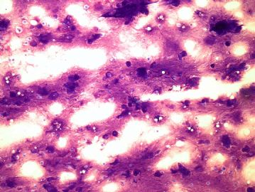
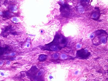
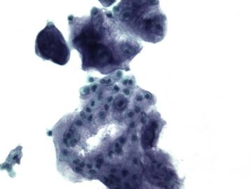
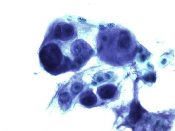
This is a FNA I did it yesterday. The patient is 70-year-old man and has a malignant tumor diagnosed in 2001. Now has a 1.5cm left chest wall subcutaneous nodule. What's your diagnosis? What's his previous malignancy?
This should be a easy one.
标签:
×参考诊断
chondrosarcoma (软骨肉瘤)
-
Most people got this one right. The patient has a history of chondrosarcoma (软骨肉瘤) and now, this is recurrent/metastatic chondrosarcoma. Just want to bring your guys attention on the diff-quick stain, which really highlights the matrix of matrix producing tumors, in this case, the rich myxoid chondroid matrix. the only time I saw a tumor other than chondrosarcoma with this much chondroid matrix was a chondroid chordoma, which is very rare. FNA of chondrosarcoma is usually easy, you see one, you will remember.


