| 图片: | |
|---|---|
| 名称: | |
| 描述: | |
- 腺癌疯了,再来一例宫颈液基
-
jiangxiaoyu 离线
- 帖子:978
- 粉蓝豆:15
- 经验:1226
- 注册时间:2007-10-31
- 加关注 | 发消息
Endometrial origin.
1. AGC vs adenocarcinoma. Pathologists should write some comment in your report or call the physician to notice your concern if you really consider ca and you want to report AGC. Anyway patients should have biopsy. In this way you do not delay the treatment of the patients.
2. Endometrial vs. endocervical: Sometimes it is very difficult to tell the origin even in the biopsy specimens. ASCCP guidline: A. Patients should have cervical and endometrial sampling (pt 35 or older) if the Pap report is AGC, endocervical origin. B. Patients should have cervical and endometrial sampling if the Pap report is AGC, endometrial origin. The origins are not very important.
3. When you have a report you always need to consider what clinician will do based on your report. We make a dx for a patient, not for a slide. Remember diagnosis and treatment.
4. Pap smears are very difficult for diagnosis. AGC is the most difficult among all abmormal Paps. You can see my online lecture powerpoint about AGC again if you are really interested in AGC. We read many Paps. I need read 60 abnormal Paps including reactive changes perday if I rotate in Pap signout week. It means 60 smears x 5 day/week (300 Pap/week). All slides were read by cytotechcicians and marked the abormal cells already. Also I know all patients's history including previous Pap, biopsy, HPV testing results. Tell you the trueth that there are many cases I really do not know how to diagnose in clinical practice. Here in the website it is more difficult to make diagnosis based on few photos (many were very poorly prepared or stained) and no any history. My feeling is like playing a game. This is why now I seldom join the discussion and diagnosis of Pap here.
Anyway good luck for your guys and patients.
| 以下是引用zhangjianxin在2008-11-25 14:01:00的发言:
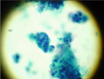
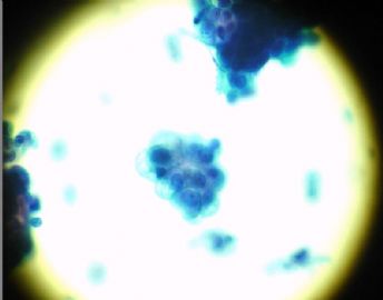
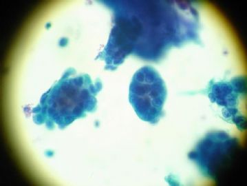
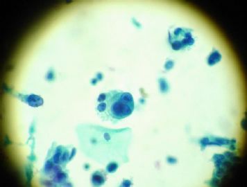
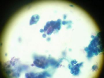
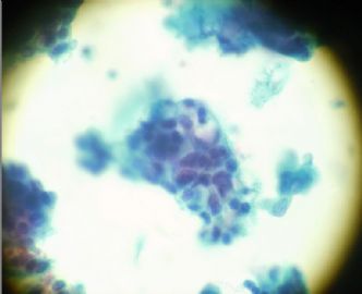
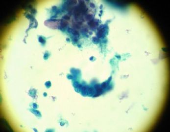
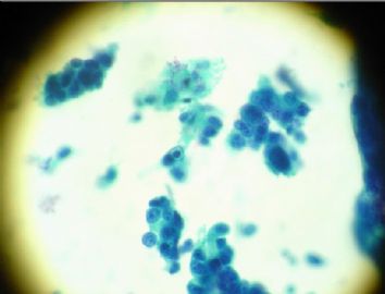
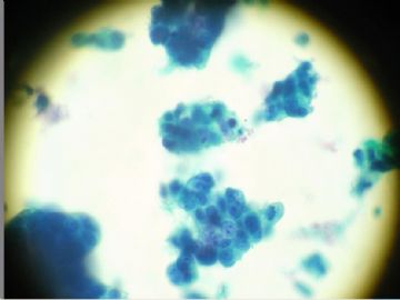
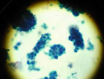
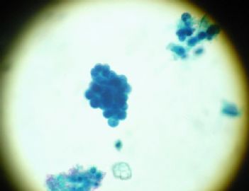
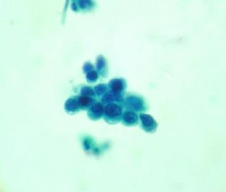
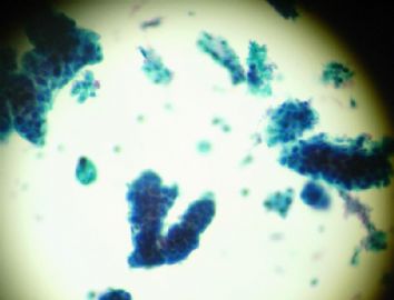
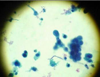
目前随着方法学及诊断水平的提高,腺上皮异常的检出比例明显增高,仅看几幅图片作判断,可能存在主观因素。从上面图片看细胞呈小圆型、偏位核、有三维结构的细胞团,倾向宫内膜来源。
子宫内膜和宫颈内膜腺癌比较
1、宫颈内膜癌比子宫内膜癌脱落细胞多
2、宫颈内膜癌比子宫内膜癌的细胞和细胞核大 3、宫颈内膜癌的核仁更多更大 4、宫颈内膜癌多为二维结构 5、宫内膜癌多为三维结构 6、随着子宫内膜癌的发展,区别二者的特征就越来越不明显
|
 学习!
学习! -
本帖最后由 于 2008-11-25 19:53:00 编辑
非典型子宫颈管细胞,倾向于肿瘤。
标准
异常胞排列呈片状、条带状、重叠。
偶见细胞团呈菊蕊团或羽毛状排列。
核增大,染色质稍增多。
偶见核分裂。
核浆比升市,胞浆量减少,细胞境界不清。
液基涂片
细胞团增厚,可呈三维结构,复层排列的细胞遮盖住团片中央部分细胞核的细节。
非典型子宫内膜细胞(图6.9-6.12)
标准
细胞团小,每团常5-10个细胞.
核与正常子宫内膜细胞相比,轻度增大.
核染色稍深.
可见小核仁.
胞浆少,偶有空泡形成.
-
zhangjianxin 离线
- 帖子:111
- 粉蓝豆:11
- 经验:111
- 注册时间:2007-03-06
- 加关注 | 发消息

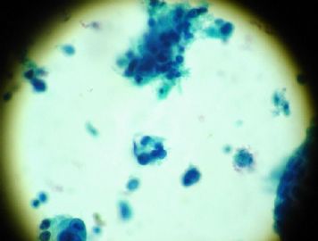
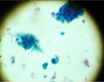
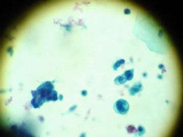
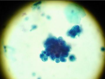


 ,错了不负责


