


| 图片: | |
|---|---|
| 名称: | |
| 描述: | |
- Lung mass FNA today Please join in the discussion
 图1
图1 图2
图2 图3
图3 图4
图4 图5
图5 图6
图6
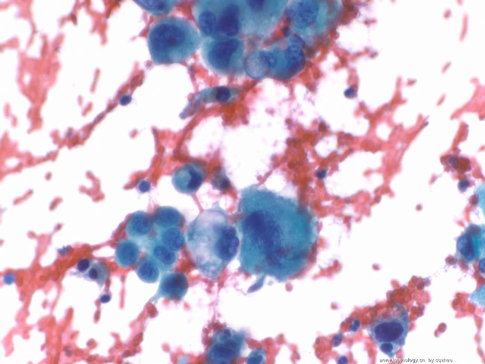
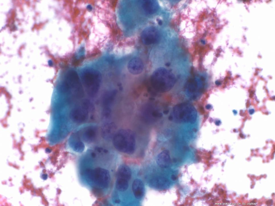
Old man with a lung mass 3 cm.
Radiologist did CT-guided FNA and I did on site evaluation this afternoon. I called malignant cells based on above one DQ. The procedure was stopped because patient had bleeding and also I think I should have enough cells for a cell block. Cytopathologists are required to give diagnosis (at least malignant, atypical, benign) on site in our institute if it is possible.
In fact I really do not know what kinds of tumor for this case. I have not seen the Pap stain yet. I have my differential dx and ordered some IHC already.
Hope people who see this case write down your differential dx and IHC.
When I have IHC results I will put here.
标签:
×参考诊断
低分化非小细胞癌
-
本帖最后由 于 2008-12-22 12:28:00 编辑
Lung mass FNA today Please join in the discussion
请大家参加本例肺穿病理讨论(cqzhao 翻译如下)Old man with a lung mass 3 cm.老男人,肺有三公分的包块, Radiologist did CT-guided FNA and I did on site evaluation this afternoon. X光大夫在CT导引下做细针肺穿操作。下午我在现场做细胞学评估, I called malignant cells based on above one DQ. 根据其中一张细胞学DQ快速染色片,我认为是恶性 The procedure was stopped because patient had bleeding因为患者穿过程中有出血and also I think I should have enough cells for a cell block. 我也认为有了足够的细胞数量了,可以做细胞块切片。因此,操作马上就停止了。 Cytopathologists are required to give diagnosis (at least malignant, atypical, benign) on site in our institute if it is possible. 我们医院规定病理学家在肺穿操作现场,其目的是尽量给出病理结论:至少要说出是否为恶性细胞,非典型细胞或良性细胞。 In fact I really do not know what kinds of tumor for this case.实际上本例我不知道这例肿瘤的结果, I have not seen the Pap stain yet.到现在也没有看巴氏涂片, I have my differential dx and ordered some IHC already.我有不同的诊断,并且要求做免疫组化, Hope people who see this case write down your differential dx and IHC.大家帮助做出诊断并且提出要做的免疫组化项目 When I have IHC results 我们的免疫组化结果出来I will put here.我马上会放到这里, Wish more people love FNA cytology希望更多的病理专家参加我们的讨论。
-
This case is interesting and may have a broad differential diagnosis. The monotonous population of cells is supporting a neoplastic process. Cells are arranged loosely with little cohesion, cells have abundant cytoplasm with cytoplasmic vaculoes. I am wondering about the possibility of metastatic Renal Cell Carcinoma? Also adrenal cortical carcinoma is in the consideration. If a young patient, germ cell tumor is in the mix? What's the history? If I have the cell block, I would do some keratin stains, S-100, LCA as my initial work-up. I look forward to hearing from Dr. Zhao.
Thank Dr. Chen's analysis. Pt has hx of ENT carcinoma. I asked the cytofellow to find surgical slides to review. My initial differeential dx includes sarcoma and carcinoma (especial RCC and squamous ca based on the hx even though the morphologic features do not look like). Some IHC stains I ordered on site have completed. I have not read them yet. I was in a shadysite cancer center for FNA last week and come back Magee this week. The fellow told me that epithelial markers are positive (PanCK, CK7), squamous markers and TTF1 are negative, vimentin and CD10 positive, EMA negative. I called the primary doctor and told him it is a carcinoma. I ordered more IHC and will review the initial stains tomorrow. Now my differential diagnoses are metastatic RCC (no any history) and metastatic poorly differential nasopharygeal ca. How do I know it is not a lung primary tumor? I will reveiw the original ENT tumor and think more how to signout the case finely.
I will like you know when I have more information.
To pathologists in China:
You see FNA cytology is interesting.
陈隆文博士译文: This case is interesting and may have a broad differential diagnosis. 本例非常有意思有许多需要鉴别,The monotonous population of cells is supporting a neoplastic process. 细胞形态比较单一,考虑为肿瘤,Cells are arranged loosely with little cohesion, cells have abundant cytoplasm with cytoplasmic vaculoes. 细胞结构松散,几乎没有一点排列,胞浆丰富,富含空泡I am wondering about the possibility of metastatic Renal Cell Carcinoma? 我有点怀疑是转移性肾细胞癌,Also adrenal cortical carcinoma is in the consideration.也考虑肾上腺皮质癌, If a young patient, germ cell tumor is in the mix? 如果患者年轻,生殖细胞肿瘤也不能完全放弃。What's the history?请详细告知病史, If I have the cell block, 如果有细胞块,I would do some keratin stains, S-100, LCA as my initial work-up. 我先要做CK,S-100,LCA。I look forward to hearing from Dr. Zhao. 我盼望着zhao大夫的最后结果。
-
CK7 positivity and EMA negative would against renal clear cell carcinoma. This is either a primary lung large cell carcinoma or a metastasis from ENT. Reviewing the slides of the patient's previous ENT tumor is the key. Thanks Dr. Zhao for the update.
| 以下是引用陈隆文博士在2008-11-26 6:42:00的发言: CK7 positivity and EMA negative would against renal clear cell carcinoma. This is either a primary lung large cell carcinoma or a metastasis from ENT. Reviewing the slides of the patient's previous ENT tumor is the key. Thanks Dr. Zhao for the update. |
试着意译,不当之处请陈博士修改,谢谢!
CK7阳性和EMA阴性针对肾透明细胞癌。这是肺大细胞癌或转移的ENT的任何一个。回顾分析病人的ENT肿瘤是关键。谢谢赵博士的更新。

- 广州金域病理
-
本帖最后由 于 2008-12-02 18:19:00 编辑
| 以下是引用cqzhao在2008-12-2 12:24:00的发言:
|
回Dr.cqzhao:如果想把更多的相片放1楼,请点编辑,继续上传就好了。
如果在回帖中放相片,先打几个字发表,然后点编辑再上传就好。
如果还不行,可能是网络问题,我发信息给坛主或管理员,请他们解决。
请Dr.cqzhao先喝杯茶,休息一下!
- 广州金域病理


