| 图片: | |
|---|---|
| 名称: | |
| 描述: | |
- Older women pelvic mesothelioma (cqz2)
-
vitamin-xbl 离线
- 帖子:383
- 粉蓝豆:0
- 经验:431
- 注册时间:2007-04-03
- 加关注 | 发消息
谢谢Dr.zhao!您的病例太精彩!并请赐教!
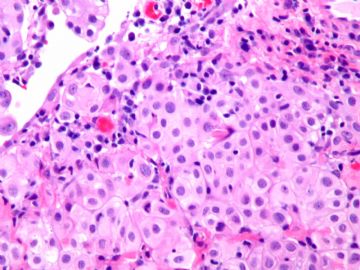
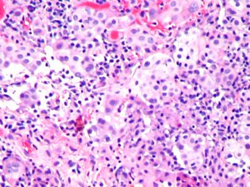
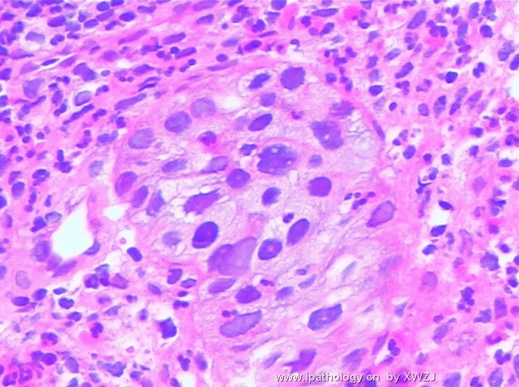
反复阅读此例,细胞圆形、立方状,浆淡红,排列呈腺泡样,细胞轻度异型,4、5楼图有乳头状的感觉,有钙化。
IHC 示:
Calretinin弥漫(+),CK5/6弥漫(+),Mesothelin弥漫(+),BerEP4(-),ER(-)支持间皮瘤的诊断。
WT1 +:肾母细胞瘤的标记,不知对间皮瘤的的诊断有何意义?
CK7+:在肺腺癌、乳腺癌、唾腺癌、胰腺癌、子宫颈管及内膜腺癌、间皮瘤,都成(+)
MOC-31 -:胆管细胞癌此抗体阳性。不知对间皮瘤有何作用?用于鉴别诊断?
综合分析诊断:盆腔间皮瘤。分化型乳头状间皮瘤?恶性间皮瘤?想请赵老师再提供较多的临床病史:肿瘤的范围等。

- 广州金域病理
-
本帖最后由 于 2008-12-27 12:27:00 编辑
| 以下是引用天山望月在2008-12-17 20:05:00的发言:
谢谢Dr.zhao!您的病例太精彩!并请赐教! 反复阅读此例,细胞圆形、立方状,浆淡红,排列呈腺泡样,细胞轻度异型,4、5楼图有乳头状的感觉,有钙化。 IHC 示: Calretinin弥漫(+),CK5/6弥漫(+),Mesothelin弥漫(+),BerEP4(-),ER(-)支持间皮瘤的诊断。 WT1 +:肾母细胞瘤的标记,不知对间皮瘤的的诊断有何意义? CK7+:在肺腺癌、乳腺癌、唾腺癌、胰腺癌、子宫颈管及内膜腺癌、间皮瘤,都成(+) MOC-31 -:胆管细胞癌此抗体阳性。不知对间皮瘤有何作用?用于鉴别诊断? 综合分析诊断:盆腔间皮瘤。分化型乳头状间皮瘤?恶性间皮瘤?想请赵老师再提供较多的临床病史:肿瘤的范围等。 |
WT1 positive for mesothelioma. It may be useful or not for differential dx. It is useful for distinction of mesothelioma from endometrioid ca or other GI tumors et al, but not useful for distinction of mesothelioma from serous papillary ca (both positive).
CK+: No use. as you mentioned mesothelial cells and many others are positive
MOC-31. it is a good epithelial marker. Almost all mesotheliomas or mesothelial cells are negative. But remember no any IHC is 100% pos or negative. I once had a case -mesothelial cells in peritoneal fluid showing strong positivity for MOC-31 fluid. This is why we often need two mesothelial markers and two epithelial markers in peritoneal/pleural cytology.
You must be or will be an excellent pathologist becuse you study so hard and carefully.Hope most pathologists in China can do the same as you did.
Thanks
-
本帖最后由 于 2008-12-22 19:19:00 编辑
31楼,大致翻译如下,不当之处请专家指导!谢谢!
CK+ :没有用。正如你所说间皮瘤细胞和其他许多肿瘤均阳性。
MOC-31:这是一个很好的上皮细胞标记。几乎所有的间皮瘤或间皮细胞是阴性的。但是,请记住没有任何免疫组化是100 %阳性或阴性。我曾经有一例腹水中的间皮细胞显示MOC-31强阳性。这就是为什么我们在腹膜/胸水细胞学检查时往往需要两个间皮标记和两个上皮标记。
您一定是或将是一个出色的病理学家,因为你学习如此的努力和细心,希望更多的中国病理学家能像你一样学习。
谢谢!

- 广州金域病理


 非常详细的解答,受益匪浅!


