共2页/25条首页上一页12下一页尾页
回复:25 阅读:7146
Liver metastatic carcinoid and differential dx (cqz 1)

cqzhao 离线

帖子:5518
粉蓝豆:1071
经验:5755
注册时间:2008-09-29
加关注  |  发消息
楼主 发表于 2008-11-17 03:50|举报|关注(1)
浏览排序[ 顺序 逆序 楼主 支持 精彩 ]  快捷回复

I see pathologists in China are not very interested in FNA. Send a cses for discussion.


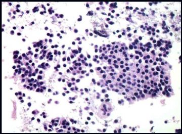
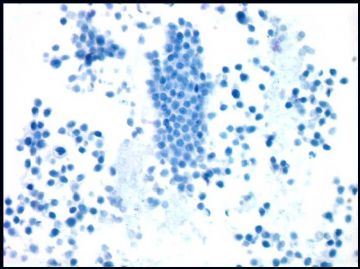
Older women with a liver mass with a history of intestinal tumor 20 y ago.

FNA of liver mass with DQ and Pap stains


(abin译:我发现国内病理医生对FNA不太感兴趣。上传一例供讨论。病史:老年女性,肝肿块,20年前有肠肿瘤病史。肝肿块FNA标本DQ染色和巴氏染色)

  • Liver metastatic carcinoid and differential dx (cqz 1)图1
    图1
  • Liver metastatic carcinoid and differential dx (cqz 1)图2
    图2
  • Liver metastatic carcinoid and differential dx (cqz 1)图3
    图3
  • Liver metastatic carcinoid and differential dx (cqz 1)图4
    图4
  • Liver metastatic carcinoid and differential dx (cqz 1)图5
    图5
标签:
本帖最后由 于 2009-03-02 07:53:00 编辑
0
×参考诊断
转移性类癌.

cqzhao 离线

帖子:5518
粉蓝豆:1071
经验:5755
注册时间:2008-09-29
加关注  |  发消息
21 楼    发表于2008-11-20 09:17:00举报|引用
返回顶部 | 快捷回复
本帖最后由 于 2008-12-10 20:19:00 编辑

Differential diagnosis鉴别诊断:

 Colon adenocarcinoma结肠腺癌

 Malignant lymphoma恶性淋巴瘤

 Small cell carcinoma小细胞癌

 Malignant Melanoma恶性黑色素瘤

 Breast carcinoma乳腺癌

 Gastric carcinoma胃癌

 Squamous cell carcinoma鳞状细胞癌

 Benign hepatocytes and bile duct epithelium良性肝细胞和胆管上皮细胞

  (天山望月译)

0
回复

cqzhao 离线

帖子:5518
粉蓝豆:1071
经验:5755
注册时间:2008-09-29
加关注  |  发消息
22 楼    发表于2008-11-20 09:15:00举报|引用
返回顶部 | 快捷回复
本帖最后由 于 2008-12-10 20:17:00 编辑

All three got correct dx or interpretation.

1. cell block

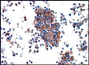
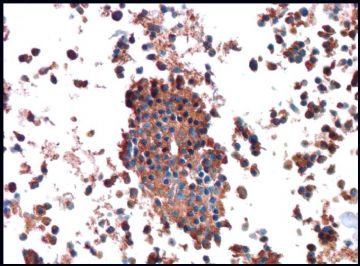
2. CD56, somatostatin, glucagon, insulin stains

3.Synaptophysin

4. chromogranin

Previous slides are not available for review. The cytologic features, IHC and history are consistent with metastatic carcinoid.


Few people are interested in FNA cytology or this case is too easy. In fact FNA cytology is the most interesting area. I will show some differential dx.

(abin译:楼上三位的诊断或解释都是对的。

1.细胞块

2.CD56、生长抑素、胰高血糖素和胰岛素染色

3.突触素

4.嗜铬素

无法获得以前的切片供复阅。细胞学特征、IHC和病史均符合转移性类癌。

很少人对FNA细胞学感兴趣或者这一例太简单了?实际上FNA细胞学是最有趣的领域。以后我会提供一些鉴别诊断。)

  • 图1
  • 图2
  • 图3
  • 图4
0
回复

wfbjwt 离线

帖子:6628
粉蓝豆:163
经验:7418
注册时间:2006-10-08
加关注  |  发消息
23 楼    发表于2008-11-17 17:15:00举报|引用
返回顶部 | 快捷回复
 类癌肝转移。需要调阅以前病理片。
0
回复
signature
嫁人就嫁灰太狼,学习要上华夏网。

天山望月 离线

帖子:4902
粉蓝豆:83
经验:5092
注册时间:2007-02-03
加关注  |  发消息
24 楼    发表于2008-11-17 17:06:00举报|引用
返回顶部 | 快捷回复

 谢谢赵老师!如此漂亮的图片!

成簇片状的小圆细胞,可见腺样和菊形团样,小圆细胞核略偏位,核膜厚,染色质粗糙,隐约可见核仁。

结合肠肿瘤病史,考虑神经内分泌癌(类癌)转移。需排除其他小细胞肿瘤。

0
回复
signature
广州金域病理

月新 离线

帖子:528
粉蓝豆:1313
经验:547
注册时间:2006-10-05
加关注  |  发消息
25 楼    发表于2008-11-17 11:24:00举报|引用
返回顶部 | 快捷回复
 感觉是神经内分泌癌。不是普通的肝细胞癌。
0
回复
回复:25 阅读:7146
共2页/25条首页上一页12下一页尾页
【免责声明】讨论内容仅作学术交流之用,不作为诊疗依据,由此而引起的法律问题作者及本站不承担任何责任。
快速回复
进入高级回复
您最多可输入10000个汉字,按 "Ctrl" + "Enter" 直接发送
搜索回复/乘电梯 ×
按内容
按会员
乘电梯
合作伙伴
友情链接