| 图片: | |
|---|---|
| 名称: | |
| 描述: | |
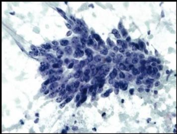
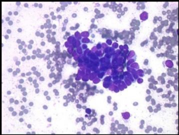
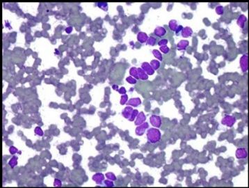
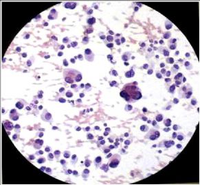
- Liver metastatic carcinoid and differential dx (cqz 1)
I see pathologists in China are not very interested in FNA. Send a cses for discussion.
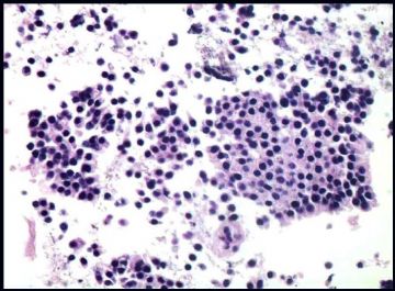
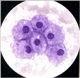
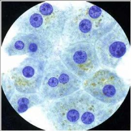
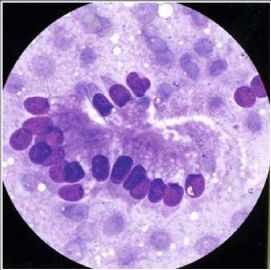
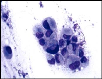
Older women with a liver mass with a history of intestinal tumor 20 y ago.
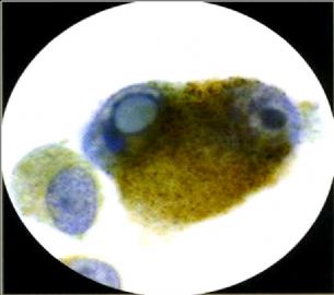
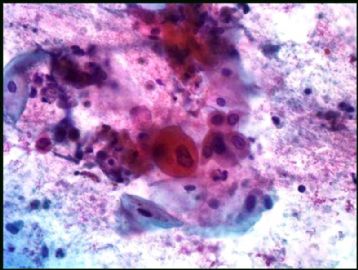
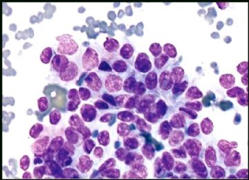
FNA of liver mass with DQ and Pap stains
(abin译:我发现国内病理医生对FNA不太感兴趣。上传一例供讨论。病史:老年女性,肝肿块,20年前有肠肿瘤病史。肝肿块FNA标本DQ染色和巴氏染色)
-
本帖最后由 于 2009-03-02 07:53:00 编辑
-
本帖最后由 于 2008-12-10 20:17:00 编辑
All three got correct dx or interpretation.
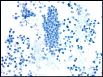
1. cell block
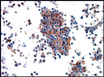
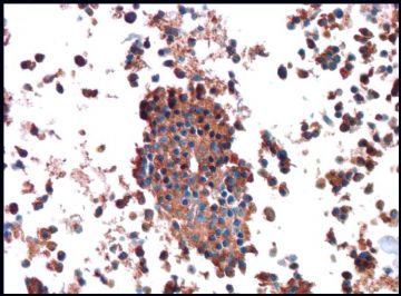
2. CD56, somatostatin, glucagon, insulin stains
3.Synaptophysin
4. chromogranin
Previous slides are not available for review. The cytologic features, IHC and history are consistent with metastatic carcinoid.
Few people are interested in FNA cytology or this case is too easy. In fact FNA cytology is the most interesting area. I will show some differential dx.
(abin译:楼上三位的诊断或解释都是对的。
1.细胞块
2.CD56、生长抑素、胰高血糖素和胰岛素染色
3.突触素
4.嗜铬素
无法获得以前的切片供复阅。细胞学特征、IHC和病史均符合转移性类癌。
很少人对FNA细胞学感兴趣或者这一例太简单了?实际上FNA细胞学是最有趣的领域。以后我会提供一些鉴别诊断。)
-
本帖最后由 于 2008-12-10 20:19:00 编辑
Differential diagnosis鉴别诊断:
Colon adenocarcinoma结肠腺癌
Malignant lymphoma恶性淋巴瘤
Small cell carcinoma小细胞癌
Malignant Melanoma恶性黑色素瘤
Breast carcinoma乳腺癌
Gastric carcinoma胃癌
Squamous cell carcinoma鳞状细胞癌
Benign hepatocytes and bile duct epithelium良性肝细胞和胆管上皮细胞
(天山望月译)
-
本帖最后由 于 2008-12-10 20:22:00 编辑
F 1-2 normal liver cells 图1-2为正常肝细胞
3. normal liver bile ductal cell 图3正常肝内胆管细胞
4. metastatic gastric ca 图4转移性胃癌
5. metastatic colonic ca 图5转移性结肠癌
6. lymphoma 图6淋巴瘤
7-8 samll cell carcinoma 图7-8小细胞癌
9-10 melanoma 图9-10恶黑
11. squamous cell ca 图11 鳞癌
(abin译)
-
本帖最后由 于 2008-12-10 20:29:00 编辑
Differential Diagnoses-Liver Metastases; (Carcinoid tumor)
鉴别诊断--类癌肝转移
nColon adenocarcinoma: Necrosis, columnar, hyperchromatic nuclei
结肠腺癌:坏死,柱状,核深染
nLymphoma: Discohesive, atypical large lymphocytes, apoptotic bodies
淋巴瘤:粘附性差,非典型性大淋巴细胞,凋亡小体
nSmall cell Carcinoma: pleomorphism, single files, nuclear molding
小细胞癌:多形性,单行排列,核镶嵌(铸型)
nMalignant melanoma: Discohesive, melanin pigment, binucleation, grooves, intranuclear pseudoinclusions
恶黑:粘附性差,黑色素,双核,核沟,核内假包涵体
nGastric carcinoma: Signet ring cells
胃癌:印戒细胞
nBreast carcinoma: Signet ring cells
乳腺癌:印戒细胞
(abin译)
Cytology- Carcinoid Tumor
n Loosely cohesive groups & single monomorphic cells
n Rosettes
n Round or plasmacytoid cells
n Round to oval nuclei
n Finely granular “salt and pepper” chromatin
n Stripped nuclei
n Basophilic cytoplasm
Intestinal Carcinoid tumor
n Low grade malignancy arising from intraepithelial endocrine stem cells in crypts.
n Most common in the ileum, also distal duodenum .
n Can metastasize to regional lymph nodes, liver, bone, skin & thyroid.
n Carcinoid syndrome: 20% with metastases, ↑ Serotonin production:
n palpitations
n cutaneous flushes
n intestinal hypermotility: vomiting &diarrhea
n Bronchospasm
n HTN
n Right valvular heart disease
Intestinal Carcinoid tumor
n Clinically diagnosis :
n plasma levels of glycoprotein chromogranin A .
n 24 hour urine levels of 5-hydroxyindoleacetic acid (5-HIAA ), a breakdown product of serotonin.
n IHC: positive for Synaptophysin, Chromogranin, CD56 +/- (most specific marker is chromogranin)
n 5 year survival: 35% with metastases, 70% localized.
Ok, hope more people like FNA cytology.
cqz
| 以下是引用cqzhao在2008-11-20 9:15:00的发言:
Previous slides are not available for review. The cytologic features, IHC and history are consistent with metastatic carcinoma.
Few people are interested in FNA cytology or this case is too easy. In fact FNA cytology is the most interesting area. I will show some differential dx. |
同感!本人对细胞学非常感兴趣!

- 广州金域病理
-
本帖最后由 于 2008-12-10 20:20:00 编辑
| 以下是引用cqzhao在2008-11-20 9:17:00的发言:
Differential diagnosis: n Colon adenocarcinoma n Malignant lymphoma n Small cell carcinoma n Malignant Melanoma n Breast carcinoma n Gastric carcinoma n Squamous cell carcinoma n Benign hepatocytes and bile duct epithelium
|
结肠腺癌
恶性淋巴瘤
小细胞癌
恶性黑色素瘤
乳腺癌
鳞状细胞癌
良性肝细胞和胆管上皮细胞

- 广州金域病理


