| 图片: | |
|---|---|
| 名称: | |
| 描述: | |
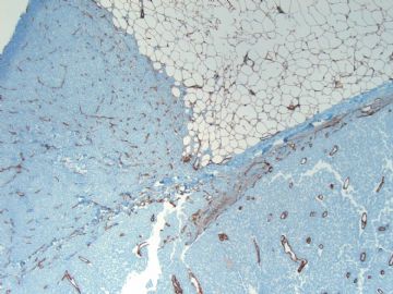
- B1728Breast encapsulated papillary carcinoma with focal frankly invasion (cqz 4)
| 姓 名: | ××× | 性别: | 年龄: | ||
| 标本名称: | |||||
| 简要病史: | |||||
| 肉眼检查: | |||||
50-55 y/f breast lesion
Fig 1-3 H&E
Fig 4 myoepithelial stain (p63)
You dx or differential dx
-
本帖最后由 于 2009-02-17 09:45:00 编辑
相关帖子
cqzhao老师的回复大意如下:
IV 胶原染色:在浸润癌部分 IV 胶原染色缺失。我很快地浏览了以下上述关于本病例的讨论,我觉得大部分都是很有道理的。
最后诊断:囊内乳头状癌(EPC,encapsulated papillary carcinoma)伴局部浸润性导管癌。照片主要显示浸润癌成分。没有显示EPC的高倍照片,故诊断有难度。我已在病例3讨论了EPC和实性乳头状癌,如果有兴趣,可以看看。
EPC可单独发生,周围乳腺组织也可伴发灶性导管原位癌(DCIS),有时可伴随显著的浸润癌成分,就如本例。大多数情况下,浸润癌为浸润性导管癌。在临床操作中关键的问题是怎样报告浸润癌的大小。大多数人认为应只报告明确的浸润癌的大小(不包括EPC部分),作为分期的依据,以避免过治疗。
本例神经内分泌染色为阴性,事实上神经内分泌阳性的肿瘤在乳腺不常见。
谢谢。
cqz
Collagen IV stain:
I quickly reviewed the discussion above. I think most comments are very reasonable for this case.
Final diangnosis: EPC with focal frankly invasive ductal carcinoma. The photos try to demostrate the EPC with focal invasion. They do not show the EPC with high power, which make the diagnosis difficult. I discussed EPC in my case 3 and solid papillary ca in other case. Please check to see the details if you want to.
EPC can be present alone or associated with focal DCIS in the surrounding breast tissue. Sometimes frankly invasive carcinoma is present in association with EPC like this case. Mostly the invasive ca is ductal ca. In clinical practice the key question is how to report the size of invasive ca. Most people think we should report only the size of the frankly invasive component (not include the EPC part) as the tumor size for staging to avoid over treatment.
Neuroendocrine stains were negative for this case. In fact neuroendocrine positive tumors are not common.
Thank for review this case.
cqz
-
本帖最后由 于 2008-11-26 23:24:00 编辑
| 以下是引用stevenshen在2008-11-26 10:08:00的发言:
Agree with Abin, in addition to invasive carcinoma, the adjacent solid component with well rounded nodule, relatively uniform cells and fibrovascular core + lack of myoepithelial staining...indicating 'solid papillary carcinoma". I have never seen a papillary solid carcinoma associated with invasive carcinoma (only heard about mucinous carcinoma association before). What about ER/PR and neuroendocrine marker profile? Beautiful case! Thanks. |
实性乳头状癌。 “我从未见过浸润性的实性乳头状癌,(以前仅仅听到关于粘液癌的) 。关于雌/孕激素受体和神经内分泌标记呢?漂亮的病例!谢谢。

- 广州金域病理
-
stevenshen 离线
- 帖子:343
- 粉蓝豆:2
- 经验:343
- 注册时间:2008-06-03
- 加关注 | 发消息
Agree with Abin, in addition to invasive carcinoma, the adjacent solid component with well rounded nodule, relatively uniform cells and fibrovascular core + lack of myoepithelial staining...indicating
'solid papillary carcinoma". I have never seen a papillary solid carcinoma associated with invasive carcinoma (only heard about mucinous carcinoma association before). What about ER/PR and neuroendocrine marker profile? Beautiful case! Thanks.
-
stevenshen 离线
- 帖子:343
- 粉蓝豆:2
- 经验:343
- 注册时间:2008-06-03
- 加关注 | 发消息


